
Saraf fasialis: Perbedaan antara revisi
| Baris 41: | Baris 41: | ||
==Gambar tambahan== |
==Gambar tambahan== |
||
<gallery> |
|||
<!-- Deleted image removed: File:Facial nerve.JPG|Facial nerve after dissection. --> |
|||
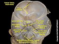
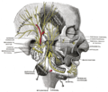
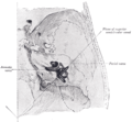
File:Cranial endobasis of a 19-20 weeks foetus.JPG|Facial nerve |
|||
File:Brain human normal inferior view with labels en.svg|Inferior view of the human brain, with the cranial nerves labelled. |
|||
Image:Gray507.png|Superficial dissection of the right side of the neck, showing the carotid and subclavian arteries. |
|||
Image:Gray567.png|Dura mater and its processes exposed by removing part of the right half of the skull, and the brain. |
|||
Image:Gray689.png|Superficial dissection of brain-stem. Ventral view. |
|||
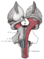
Image:Gray719.png|Hind- and mid-brains; postero-lateral view. |
|||
Image:Gray780.png|The sphenopalatine ganglion and its branches. |
|||
Image:Gray781.png|Mandibular division of the trifacial nerve. |
|||
Image:Gray782.png|Mandibular division of trifacial nerve, seen from the middle line. |
|||
Image:Gray788.png|Plan of the facial and intermediate nerves and their communication with other nerves. |
|||
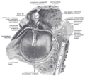
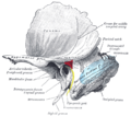
Image:Gray789.png |The course and connections of the facial nerve in the [[temporal bone]]. |
|||
Image:Gray792.png|Upper part of medulla spinalis and hind- and mid-brains; posterior aspect, exposed in situ. |
|||
Image:Gray911.png|View of the inner wall of the tympanum (enlarged.) |
|||
Image:Gray912.png|The right membrana tympani with the hammer and the chorda tympani, viewed from within, from behind, and from above. |
|||
Image:Gray922.png|Position of the right bony labyrinth of the ear in the skull, viewed from above. |
|||
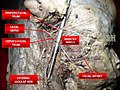
Image:Facial nerve dissected.jpg|Facial nerve dissected |
|||
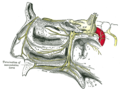
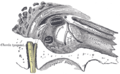
Image:Gray1209.png|Left temporal bone showing surface markings for the tympanic antrum (red), transverse sinus (blue), and facial nerve (yellow). |
|||
Image:Gray1210.png|Side of neck, showing chief surface markings. |
|||
<!-- Missing image removed: Image:Illu cranial nerves.jpg|Cranial nerves --> |
|||
Image:Head facial nerve branches.jpg|Head facial nerve branches |
|||
File:Facial nerve at foetus 1.jpg|Facial nerve at foetus |
|||
File:Facial nerve at foetus 2.jpg|Facial nerve at foetus |
|||
File:Facial nerve at foetus 3.jpg|Facial nerve at foetus |
|||
File:Facial nerve at foetus 4.jpg|Facial nerve at foetus |
|||
Image:Facial canal.png|Facial canal |
|||
File:Slide1rrrr.JPG|Facial nerve |
|||
</gallery> |
|||
==Lihat pula== |
==Lihat pula== |
||
Revisi per 8 Agustus 2012 04.09
Artikel ini memberikan informasi dasar tentang topik kesehatan. |
| Saraf fasialis | ||
|---|---|---|
 | ||
| Cranial nerve VII | ||
 | ||
| The nerves of the scalp, face, and side of neck. | ||
| Latin | nervus facialis | |
| Persarafan | ||
| Asal | ||
| Ke | ||
| Saraf Kranial |
|---|
| CN I – Olfaktorius |
| CN II – Optikus |
| CN III – Okulomotor |
| CN IV – Troklearis |
| CN V – Trigeminus |
| CN VI – Abdusen |
| CN VII – Fasialis |
| CN VIII – Vestibulokoklearis |
| CN IX – Glosofaringeal |
| CN X – Vagus |
| CN XI – Aksesorius |
| CN XII – Hipoglossus |
Saraf fasialis adalah saraf kranialis ke-7 berperan besar dalam mengatur ekspresi dan indra perasa di kulit wajah manusia. Saraf fasialis memiliki 2 komponen utama. Komponen yang lebih besar merupakan murni saraf motorik dan berperan dalam persarafan otot ekspresi wajah. Komponen ini yang merupakan saraf fasialis sesungguhnya. Akan tetapi sepanjang perjalanan komponen besar terdapat komponen yang lebih tipis yang disebut saraf intermedius. Saraf intermedius mengandung serabut saraf viseral dan serabut aferen somatis.[1]
Fungsi
Saraf fasialis utamanya berperan dalam memasok impuls untuk otot-otot ekspresi wajah. Disamping itu saraf fasialis juga berfungsi sebagai: • Penyalur sensasi dari bagian anterior lidah dan rongga mulut • Melalui persarafan parasimpatis saraf facialis, kelenjar saliva,lakrimal, hidung dan kelenjar palatina bisa menghasilkan sekret [2]
Letak
Saraf fasialis berasal dari sudut cerebellopontine - bagian lateral dari persimpangan pontomedullary • Memiliki dua akar saraf yang berdekatan yakni motor root (lebih besar, lebih medial)dan saraf intermedius (lebih kecil, lebih lateral) - disebut saraf intermedius karena ditemukan diantara dua saraf yang lebih besar (akar utama VII dan VIII). Nervus intermedius memiliki serat parasimpatis dan sensorik dan yang awalnya merupakan bagian dari saraf VIII.[2]
Kelainan Saraf Fasialis
- Penyakit Parotis
Tumor parotis, trauma atau operasi parotis dapat merusak cabang dari saraf fasialis. Hal ini akan mengakibatkan palsy wajah ipsilateral(satu sisi) dan kehilangan fungsi fungsionalnya. Sejauh ini tidak ada pasien yang dapat pulih sempurna dari kondisi ini.
- Gangguang pada otot Stapedius: hyperacusis[2]
Disfungsi dari otot terkecil diakibatkan oleh saraf fasialis dapat menyebabkan gejala yang menyedihkan. Otot stapedius mengatur gerakan dari rantai tulang pendengaran dan jika tidak aktif, suara akan menyimpang dan bergema yang diswebut kelainan hyperacusis[2]
- Bell palsy
Merupakan kelainan yang sering dijumpai akibat kerusakan saraf fasialis, biasa disebut facial palsy. Etiologi sebenarnya hingga kini masih belum diketahui secara pasti. Akan tetapi beberapa faktor seperti spasme pembuluh darah arteri di kanal wajah yang memasok nutrisi dari saraf fasialis ataupun peradangan dan pembengkakan saraf dalam kanal tulang kemungkinan bertanggung jawab terhadap kondisi ini.[2]
Gambar tambahan
-
 Facial nerve
 -
 Inferior view of the human brain, with the cranial nerves labelled.
 -
 Superficial dissection of the right side of the neck, showing the carotid and subclavian arteries.
 -
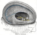 Dura mater and its processes exposed by removing part of the right half of the skull, and the brain.
 -
 Superficial dissection of brain-stem. Ventral view.
 -
 Hind- and mid-brains; postero-lateral view.
 -
 The sphenopalatine ganglion and its branches.
 -
 Mandibular division of the trifacial nerve.
 -
 Mandibular division of trifacial nerve, seen from the middle line.
 -
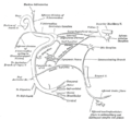 Plan of the facial and intermediate nerves and their communication with other nerves.
 -
 The course and connections of the facial nerve in the temporal bone.
 -
 Upper part of medulla spinalis and hind- and mid-brains; posterior aspect, exposed in situ.
 -
 View of the inner wall of the tympanum (enlarged.)
 -
 The right membrana tympani with the hammer and the chorda tympani, viewed from within, from behind, and from above.
 -
 Position of the right bony labyrinth of the ear in the skull, viewed from above.
 -
 Facial nerve dissected
 -
 Left temporal bone showing surface markings for the tympanic antrum (red), transverse sinus (blue), and facial nerve (yellow).
 -
 Side of neck, showing chief surface markings.
 -
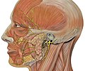 Head facial nerve branches
 -
 Facial nerve at foetus
 -
 Facial nerve at foetus
 -
 Facial nerve at foetus
 -
 Facial nerve at foetus
 -
 Facial canal
 -
 Facial nerve


Lihat pula
Referensi
Pranala luar
- (Inggris) ent/8 di eMedicine
- (Inggris) position of facial nerve on MRI
- (Inggris) WUSTL - map
- (Inggris) Notes on Facial Nerve