Melioidosis: Perbedaan antara revisi
Hanamanteo (bicara | kontrib) + Tag: halaman dengan galat kutipan |
Hanamanteo (bicara | kontrib) + Tag: halaman dengan galat kutipan |
||
| Baris 27: | Baris 27: | ||
'''Melioidosis''' adalah penyakit [[infeksi]] yang disebabkan oleh [[bakteri]] [[Gram-negatif]] bernama ''[[Burkholderia pseudomallei]]''.<ref name="Joost 2018"/> Kebanyakan orang yang dijangkiti ''Burkholderia pseudomallei'' tidak mengalami satupun gejala, tetapi mereka yang mengalami gejala memiliki tanda dan gejala dari gejala ringan seperti [[demam]], perubahan kulit, [[radang paru-paru]], dan [[bisul]], hingga gejala berat seperti [[ensefalomielitis|radang otak]], [[radang sendi]], dan [[kejang septik|tekanan darah rendah yang berbahaya]] yang menyebabkan kematian.<ref name="Joost 2018"/> Sekitar 10% dari orang penderita melioidosis mengalami gejala yang berlangsung lebih dari dua bulan yang disebut melioidosis kronis.<ref name="Joost 2018"/> |
'''Melioidosis''' adalah penyakit [[infeksi]] yang disebabkan oleh [[bakteri]] [[Gram-negatif]] bernama ''[[Burkholderia pseudomallei]]''.<ref name="Joost 2018"/> Kebanyakan orang yang dijangkiti ''Burkholderia pseudomallei'' tidak mengalami satupun gejala, tetapi mereka yang mengalami gejala memiliki tanda dan gejala dari gejala ringan seperti [[demam]], perubahan kulit, [[radang paru-paru]], dan [[bisul]], hingga gejala berat seperti [[ensefalomielitis|radang otak]], [[radang sendi]], dan [[kejang septik|tekanan darah rendah yang berbahaya]] yang menyebabkan kematian.<ref name="Joost 2018"/> Sekitar 10% dari orang penderita melioidosis mengalami gejala yang berlangsung lebih dari dua bulan yang disebut melioidosis kronis.<ref name="Joost 2018"/> |
||
Manusia dijangkiti ''Burkholderia pseudomallei'' melalui kontak dengan air yang tercemar. Bakteri ini masuk ke dalam tubuh melalui luka, tarikan napas, atau penelanan. Penularan dari manusia ke manusia atau dari hewan ke manusia sangat jarang terjadi.<ref name="Joost 2018"/> Infeksi ini [[endemi (epidemiologi)|masih ada]] di Asia Tenggara, khususnya di timur laut [[Thailand]] dan utara [[Australia]].<ref name="Joost 2018"/> Di negara-negara maju seperti Eropa dan Amerika Serikat, kasus melioidosis umumnya diimpor dari negara-negara tempat melioidosis lebih sering terjadi.<ref name="Currie 2015"/> Tanda dan gejala melioidosis menyerupai [[tuberkulosis]] dan sering terjadi kesalahan diagnosis.<ref>{{Cite journal|last1=Brightman|first1=Christopher|last2=Locum|date=2020|title=Melioidosis: the Vietnamese time bomb|journal=Trends in Urology & Men's Health|language=en|volume=11|issue=3|pages=30–32|doi=10.1002/tre.753|issn=2044-3749|doi-access=free}}</ref><ref name="Yi 2014"/> Diagnosis biasanya dikonfirmasi oleh pertumbuhan ''Burkholderia pseudomallei'' dari darah atau cairan tubuh orang yang dijangkiti lainnya.<ref name="Joost 2018"/> Mereka yang menderita melioidosis pertama-tama diobati dengan antibiotik intravena "fase intensif" (paling sering [[seftazidima]]) diikuti dengan pengobatan [[kotrimoksazol]] selama beberapa bulan.<ref name="Joost 2018"/> |
Manusia dijangkiti ''Burkholderia pseudomallei'' melalui kontak dengan air yang tercemar. Bakteri ini masuk ke dalam tubuh melalui luka, tarikan napas, atau penelanan. Penularan dari manusia ke manusia atau dari hewan ke manusia sangat jarang terjadi.<ref name="Joost 2018"/> Infeksi ini [[endemi (epidemiologi)|masih ada]] di Asia Tenggara, khususnya di timur laut [[Thailand]] dan utara [[Australia]].<ref name="Joost 2018"/> Di negara-negara maju seperti Eropa dan Amerika Serikat, kasus melioidosis umumnya diimpor dari negara-negara tempat melioidosis lebih sering terjadi.<ref name="Currie 2015"/> Tanda dan gejala melioidosis menyerupai [[tuberkulosis]] dan sering terjadi kesalahan diagnosis.<ref>{{Cite journal|last1=Brightman|first1=Christopher|last2=Locum|date=2020|title=Melioidosis: the Vietnamese time bomb|journal=Trends in Urology & Men's Health|language=en|volume=11|issue=3|pages=30–32|doi=10.1002/tre.753|issn=2044-3749|doi-access=free}}</ref><ref name="Yi 2014"/> Diagnosis biasanya dikonfirmasi oleh pertumbuhan ''Burkholderia pseudomallei'' dari darah atau cairan tubuh orang yang dijangkiti lainnya.<ref name="Joost 2018"/> Mereka yang menderita melioidosis pertama-tama diobati dengan antibiotik intravena "fase intensif" (paling sering [[seftazidima]]) diikuti dengan pengobatan [[Trimetoprim/sulfametoksazol|kotrimoksazol]] selama beberapa bulan.<ref name="Joost 2018"/> Bahkan jika dirawat dengan cermat, sekitar 10% penderita melioidosis meninggal karenanya. Jika tidak ditangani dengan cermat, tingkat kematian bisa melonjak hingga 40%.<ref name="Joost 2018"/> |
||
Upaya pencegahan melioidosis antara lain memakai alat pelindung diri saat menangani air yang terkontaminasi, membiasakan kebersihan tangan, minum air matang, dan menghindari kontak langsung dengan tanah, air, atau hujan lebat. [[Antibiotik]] kotrimoksazol hanya digunakan sebagai pencegahan untuk individu yang berisiko tinggi terkena melioidosis setelah terpapar bakteri. Tiada vaksin untuk melioidosis yang telah disetujui.<ref name="Joost 2018"/> |
|||
Sekitar 165 ribu orang dijangkiti melioidosis tiap tahun dan menewaskan 89 ribu orang. [[Diabetes melitus|Diabetes]] adalah faktor risiko utama penyakit melioidosis dengan lebih dari setengah kasus melioidosis terjadi pada penderita diabetes.<ref name="Joost 2018"/> Peningkatan curah hujan dikaitkan dengan lonjakan jumlah kasus melioidosis di daerah endemi.<ref name="Yi 2014"/> Melioidosis pertama kali dideskripsikan oleh [[Alfred Whitmore]] pada tahun 1912 di wilayah yang saat ini bernama [[Myanmar]].<ref name="Whitmore 1912"/> |
|||
== Tanda dan gejala == |
|||
=== Akut === |
|||
[[Berkas:Signs of melioidosis.svg|thumb|upright=1.3|Schematic depiction of the signs of melioidosis]] |
|||
[[Berkas:Melioidosis PA and lateral X rays.jpg|thumb|upright=1.3|Chest X-ray showing opacity of the left middle and lower zones of the lung.]] |
|||
[[Berkas:CT and MRI scan of the brain with melioidosis.jpg|thumb|upright=1.3|CT and MRI scans showing lesion of the right frontal lobe of the brain.]] |
|||
[[Berkas:Septic arthritis of left hip joint with melioidosis.jpg|thumb|upright=1.3|Septic arthritis of the left hip with joint destruction]] |
|||
Pajanan terhadap ''Burkholderia pseudomallei'' biasanya dapat menyebabkan antibodi diproduksi untuk melawan bakteri itu tanpa gejala apapun. Dari pasien yang menderita infeksi klinis, 85% pasien mengalami gejala akut dari pemerolehan bakteri terkini.<ref name="Joost 2018"/><ref name="pmid21152057">{{cite journal| author=Currie BJ, Ward L, Cheng AC| title=The epidemiology and clinical spectrum of melioidosis: 540 cases from the 20 year Darwin prospective study. | journal=PLOS Negl Trop Dis | year= 2010 | volume= 4 | issue= 11 | pages= e900 | pmid=21152057 | doi=10.1371/journal.pntd.0000900 | pmc=2994918 }}</ref><ref name="Bennett 2015">{{cite book |vauthors=Bennett JE, Raphael D, Martin JB, Currie BJ |title=Mandell, Douglas, and Bennett's Principles and Practice of Infectious Diseases|chapter=223 |date=2015 |publisher=Elsevier |isbn=978-1-4557-4801-3 |pages=2541–2549 |edition=Eighth}}</ref> [[Masa inkubasi]] rata-rata melioidosis akut adalah 9 hari (kisaran 1–21 hari).<ref name="Joost 2018"/> Nevertheless, symptoms of melioidosis can appear in 24 hours for those who are infected during a near drowning in contaminated water.<ref name="Bennett 2015"/> Those affected present with symptoms of [[sepsis]] (predominantly fever) with or without [[pneumonia]], or localised [[abscess]] or other focus of infection. The presence of nonspecific signs and symptoms has caused melioidosis to be nicknamed "the great mimicker".<ref name="Joost 2018"/> |
|||
People with [[diabetes mellitus]] or regular exposure to the bacteria are at increased risk of developing melioidosis. The disease should be considered in those staying in endemic areas who develop fever, pneumonia, or abscesses in their liver, spleen, prostate, or [[parotid gland]]s.<ref name="Joost 2018"/> The clinical manifestation of the disease can range from simple skin changes to severe organ problems.<ref name="Joost 2018"/> Skin changes can be nonspecific abscesses or ulcerations.<ref>{{cite journal | vauthors = Fertitta L, Monsel G, Torresi J, Caumes E | title = Cutaneous melioidosis: a review of the literature | journal = International Journal of Dermatology | volume = 58 | issue = 2 | pages = 221–227 | date = February 2019 | pmid = 30132827 | doi = 10.1111/ijd.14167 | s2cid = 52056443 | hdl = 11343/284394 | hdl-access = free }}</ref> In northern Australia, 60% of the infected children presented with only skin lesions, while 20% presented with pneumonia.<ref name="Currie 2015"/> The commonest organs affected are liver, spleen, lungs, prostate, and kidneys. Among the most common clinical signs are [[bacteremia|presence of bacteria in blood]] (in 40 to 60% of cases), pneumonia (50%), and [[septic shock]] (20%).<ref name="Joost 2018"/> People with only pneumonia may have a prominent cough with sputum and shortness of breath. However, those with septic shock together with pneumonia may have minimal coughing.<ref name="Yi 2014"/> Results of a chest X-ray can range from diffuse nodular infiltrates in those with septic shock to progressive [[pulmonary consolidation|solidification of the lungs]] in the [[Lung#Anatomy|upper lobes]] for those with pneumonia only. [[Pleural effusion|Excess fluid in the pleural cavity]] and [[empyema|gathering of pus within a cavity]] are more common for melioidosis affecting lower lobes of the lungs.<ref name="Yi 2014"/> In 10% of cases, people develop secondary pneumonia caused by other bacteria after the primary infection.<ref name="Currie 2015"/> |
|||
Depending on the course of infection, other severe manifestations develop. About 1 to 5% of those infected develop [[encephalomyelitis|inflammation of the brain and brain covering]] or [[brain abscess|collection of pus in the brain]]; 14 to 28% develop [[acute pyelonephritis|bacterial inflammation of the kidneys]], kidney abscess or [[prostatic abscess]]es; 0 to 30% develop neck or [[parotid gland|salivary gland]] abscesses; 10 to 33% develop liver, spleen, or paraintestinal abscesses; 4 to 14% develop [[septic arthritis]] and [[osteomyelitis]].<ref name="Joost 2018"/> Rare manifestations include [[lymphadenopathy|lymph node disease]] resembling tuberculosis,<ref name="Gassiep 2020">{{cite journal | vauthors = Gassiep I, Armstrong M, Norton R | title = Human Melioidosis | journal = Clinical Microbiology Reviews | volume = 33 | issue = 2 | date = March 2020 | pmid = 32161067 | pmc = 7067580 | doi = 10.1128/CMR.00006-19 }}</ref> [[mediastinum|mediastinal]] masses, [[pericardial effusion|collection of fluid in the heart covering]],<ref name="Currie 2015"/> [[mycotic aneurysm|abnormal dilatation of blood vessels due to infection]],<ref name="Joost 2018"/> and [[pancreatitis|inflammation of the pancreas]].<ref name="Currie 2015"/> In Australia, up to 20% of infected males develop prostatic abscess characterized by [[dysuria|pain during urination]], difficulty in passing urine, and [[urinary retention]] requiring [[catheter]]isation.<ref name="Joost 2018"/> [[Rectal examination]] shows inflammation of the [[prostate]].<ref name="Currie 2015"/> In Thailand, 30% of the infected children develop parotid abscesses.<ref name="Joost 2018"/> Encephalomyelitis can occur in healthy people without risk factors. Those with melioidosis encephomyelitis tend to have normal [[computed tomography]] scans, but increased [[T2*-weighted imaging|T2 signal]] by [[magnetic resonance imaging]], extending to the [[brain stem]] and [[spinal cord]]. Clinical signs include: unilateral [[upper motor neuron]] limb weakness, [[focal neurological signs|cerebellar signs]], and cranial nerve palsies ([[Sixth nerve palsy|VI]], [[Facial nerve paralysis|VII]] nerve palsies and [[bulbar palsy]]). Some cases presented with [[flaccid paralysis]] alone.<ref name="Currie 2015"/> In northern Australia, all melioidosis with encephalomyelitis cases had elevated white cells in the [[cerebrospinal fluid]] (CSF), mostly [[mononuclear cell]]s with elevated CSF protein.<ref name="Gassiep 2020"/> |
|||
== Referensi == |
== Referensi == |
||
Revisi per 29 Oktober 2021 07.02
Halaman ini sedang dipersiapkan dan dikembangkan sehingga mungkin terjadi perubahan besar. Anda dapat membantu dalam penyuntingan halaman ini. Halaman ini terakhir disunting oleh Hanamanteo (Kontrib • Log) 911 hari 1398 menit lalu. Jika Anda melihat halaman ini tidak disunting dalam beberapa hari, mohon hapus templat ini. |
| Melioidosis | |
|---|---|
 | |
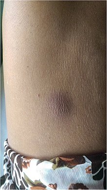
| Bisul melioidosis di perut | |
| Informasi umum | |
| Spesialisasi | Penyakit menular |
| Penyebab | Burkholderia pseudomallei spread by contact to soil or water[1] |
| Faktor risiko | Diabetes mellitus, thalassaemia, alcoholism, chronic kidney disease[1] |
| Aspek klinis | |
| Gejala dan tanda | Tiada, demam, radang paru-paru, beberapa bisul[1] |
| Komplikasi | Encephalomyelitis, septic shock, acute pyelonephritis, septic arthritis, osteomyelitis[1] |
| Awal muncul | 1-21 hari setelah terjangkit[1] |
| Diagnosis | Mengembangkan bakteri di perantara kultur[1] |
| Kondisi serupa | Tuberculosis[2] |
| Tata laksana | |
| Pencegahan | Mencegah dari kontak dengan air yang terkontaminasi, profilaksis antibiotik[1] |
| Perawatan | Ceftazidime, meropenem, co-trimoxazole[1] |
| Distribusi dan frekuensi | |
| Prevalensi | 165,000 orang tiap tahun[1] |
| Kematian | 89,000 orang tiap tahunr[1] |
Melioidosis adalah penyakit infeksi yang disebabkan oleh bakteri Gram-negatif bernama Burkholderia pseudomallei.[1] Kebanyakan orang yang dijangkiti Burkholderia pseudomallei tidak mengalami satupun gejala, tetapi mereka yang mengalami gejala memiliki tanda dan gejala dari gejala ringan seperti demam, perubahan kulit, radang paru-paru, dan bisul, hingga gejala berat seperti radang otak, radang sendi, dan tekanan darah rendah yang berbahaya yang menyebabkan kematian.[1] Sekitar 10% dari orang penderita melioidosis mengalami gejala yang berlangsung lebih dari dua bulan yang disebut melioidosis kronis.[1]
Manusia dijangkiti Burkholderia pseudomallei melalui kontak dengan air yang tercemar. Bakteri ini masuk ke dalam tubuh melalui luka, tarikan napas, atau penelanan. Penularan dari manusia ke manusia atau dari hewan ke manusia sangat jarang terjadi.[1] Infeksi ini masih ada di Asia Tenggara, khususnya di timur laut Thailand dan utara Australia.[1] Di negara-negara maju seperti Eropa dan Amerika Serikat, kasus melioidosis umumnya diimpor dari negara-negara tempat melioidosis lebih sering terjadi.[3] Tanda dan gejala melioidosis menyerupai tuberkulosis dan sering terjadi kesalahan diagnosis.[4][2] Diagnosis biasanya dikonfirmasi oleh pertumbuhan Burkholderia pseudomallei dari darah atau cairan tubuh orang yang dijangkiti lainnya.[1] Mereka yang menderita melioidosis pertama-tama diobati dengan antibiotik intravena "fase intensif" (paling sering seftazidima) diikuti dengan pengobatan kotrimoksazol selama beberapa bulan.[1] Bahkan jika dirawat dengan cermat, sekitar 10% penderita melioidosis meninggal karenanya. Jika tidak ditangani dengan cermat, tingkat kematian bisa melonjak hingga 40%.[1]
Upaya pencegahan melioidosis antara lain memakai alat pelindung diri saat menangani air yang terkontaminasi, membiasakan kebersihan tangan, minum air matang, dan menghindari kontak langsung dengan tanah, air, atau hujan lebat. Antibiotik kotrimoksazol hanya digunakan sebagai pencegahan untuk individu yang berisiko tinggi terkena melioidosis setelah terpapar bakteri. Tiada vaksin untuk melioidosis yang telah disetujui.[1]
Sekitar 165 ribu orang dijangkiti melioidosis tiap tahun dan menewaskan 89 ribu orang. Diabetes adalah faktor risiko utama penyakit melioidosis dengan lebih dari setengah kasus melioidosis terjadi pada penderita diabetes.[1] Peningkatan curah hujan dikaitkan dengan lonjakan jumlah kasus melioidosis di daerah endemi.[2] Melioidosis pertama kali dideskripsikan oleh Alfred Whitmore pada tahun 1912 di wilayah yang saat ini bernama Myanmar.[5]
Tanda dan gejala
Akut


Pajanan terhadap Burkholderia pseudomallei biasanya dapat menyebabkan antibodi diproduksi untuk melawan bakteri itu tanpa gejala apapun. Dari pasien yang menderita infeksi klinis, 85% pasien mengalami gejala akut dari pemerolehan bakteri terkini.[1][6][7] Masa inkubasi rata-rata melioidosis akut adalah 9 hari (kisaran 1–21 hari).[1] Nevertheless, symptoms of melioidosis can appear in 24 hours for those who are infected during a near drowning in contaminated water.[7] Those affected present with symptoms of sepsis (predominantly fever) with or without pneumonia, or localised abscess or other focus of infection. The presence of nonspecific signs and symptoms has caused melioidosis to be nicknamed "the great mimicker".[1]
People with diabetes mellitus or regular exposure to the bacteria are at increased risk of developing melioidosis. The disease should be considered in those staying in endemic areas who develop fever, pneumonia, or abscesses in their liver, spleen, prostate, or parotid glands.[1] The clinical manifestation of the disease can range from simple skin changes to severe organ problems.[1] Skin changes can be nonspecific abscesses or ulcerations.[8] In northern Australia, 60% of the infected children presented with only skin lesions, while 20% presented with pneumonia.[3] The commonest organs affected are liver, spleen, lungs, prostate, and kidneys. Among the most common clinical signs are presence of bacteria in blood (in 40 to 60% of cases), pneumonia (50%), and septic shock (20%).[1] People with only pneumonia may have a prominent cough with sputum and shortness of breath. However, those with septic shock together with pneumonia may have minimal coughing.[2] Results of a chest X-ray can range from diffuse nodular infiltrates in those with septic shock to progressive solidification of the lungs in the upper lobes for those with pneumonia only. Excess fluid in the pleural cavity and gathering of pus within a cavity are more common for melioidosis affecting lower lobes of the lungs.[2] In 10% of cases, people develop secondary pneumonia caused by other bacteria after the primary infection.[3]
Depending on the course of infection, other severe manifestations develop. About 1 to 5% of those infected develop inflammation of the brain and brain covering or collection of pus in the brain; 14 to 28% develop bacterial inflammation of the kidneys, kidney abscess or prostatic abscesses; 0 to 30% develop neck or salivary gland abscesses; 10 to 33% develop liver, spleen, or paraintestinal abscesses; 4 to 14% develop septic arthritis and osteomyelitis.[1] Rare manifestations include lymph node disease resembling tuberculosis,[9] mediastinal masses, collection of fluid in the heart covering,[3] abnormal dilatation of blood vessels due to infection,[1] and inflammation of the pancreas.[3] In Australia, up to 20% of infected males develop prostatic abscess characterized by pain during urination, difficulty in passing urine, and urinary retention requiring catheterisation.[1] Rectal examination shows inflammation of the prostate.[3] In Thailand, 30% of the infected children develop parotid abscesses.[1] Encephalomyelitis can occur in healthy people without risk factors. Those with melioidosis encephomyelitis tend to have normal computed tomography scans, but increased T2 signal by magnetic resonance imaging, extending to the brain stem and spinal cord. Clinical signs include: unilateral upper motor neuron limb weakness, cerebellar signs, and cranial nerve palsies (VI, VII nerve palsies and bulbar palsy). Some cases presented with flaccid paralysis alone.[3] In northern Australia, all melioidosis with encephalomyelitis cases had elevated white cells in the cerebrospinal fluid (CSF), mostly mononuclear cells with elevated CSF protein.[9]
Referensi
- ^ a b c d e f g h i j k l m n o p q r s t u v w x y z aa ab ac ad Kesalahan pengutipan: Tag
<ref>tidak sah; tidak ditemukan teks untuk ref bernamaJoost 2018 - ^ a b c d e Kesalahan pengutipan: Tag
<ref>tidak sah; tidak ditemukan teks untuk ref bernamaYi 2014 - ^ a b c d e f g Kesalahan pengutipan: Tag
<ref>tidak sah; tidak ditemukan teks untuk ref bernamaCurrie 2015 - ^ Brightman, Christopher; Locum (2020). "Melioidosis: the Vietnamese time bomb". Trends in Urology & Men's Health (dalam bahasa Inggris). 11 (3): 30–32. doi:10.1002/tre.753
 . ISSN 2044-3749.
- ^ Kesalahan pengutipan: Tag
<ref>tidak sah; tidak ditemukan teks untuk ref bernamaWhitmore 1912 - ^ Currie BJ, Ward L, Cheng AC (2010). "The epidemiology and clinical spectrum of melioidosis: 540 cases from the 20 year Darwin prospective study". PLOS Negl Trop Dis. 4 (11): e900. doi:10.1371/journal.pntd.0000900. PMC 2994918
 . PMID 21152057.
- ^ a b Bennett JE, Raphael D, Martin JB, Currie BJ (2015). "223". Mandell, Douglas, and Bennett's Principles and Practice of Infectious Diseases (edisi ke-Eighth). Elsevier. hlm. 2541–2549. ISBN 978-1-4557-4801-3.
- ^ Fertitta L, Monsel G, Torresi J, Caumes E (February 2019). "Cutaneous melioidosis: a review of the literature". International Journal of Dermatology. 58 (2): 221–227. doi:10.1111/ijd.14167. hdl:11343/284394
 . PMID 30132827.
- ^ a b Gassiep I, Armstrong M, Norton R (March 2020). "Human Melioidosis". Clinical Microbiology Reviews. 33 (2). doi:10.1128/CMR.00006-19. PMC 7067580
 . PMID 32161067.
Pranala luar
| Klasifikasi | |
|---|---|
| Sumber luar |
- Resource Center for melioidosis
- Templat:CDCDiseaseInfo
- Burkholderia pseudomallei genomes and related information at PATRIC, a Bioinformatics Resource Center funded by NIAID
- Monograph on Melioidosis (ISBN 978-0-444-53479-8); Elsevier Press, 2012, https://www.researchgate.net/publication/354857974_Monograph_Melioidosis-a-century-of-observation-and-research_ISBN_978-0-444-53479-8
